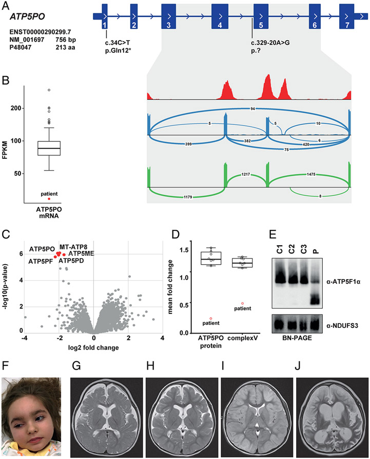
Figure 3: Biallelic ATP5PO variants identified in 1 case.
(a) RNA-seq-based detection of a splice defect induced by the intronic variant c.329-20A>G. Sashimi plots to visualize splice junctions demonstrate that the c.329-20A>G allele causes skipping of exon 5 and exons 4 plus 5 (blue graph), resulting in reduced total amounts of ATP5PO mRNA levels (b). Wild-type ATP5PO produces only the normal transcript (green graph). (c) Proteomic analysis of patient-derived fibroblasts shows significant down-regulation of five ATPase subunits (red dots) relative to controls. (d) Mass spectrometry-based protein quantification demonstrates a decrease of ATP5PO protein and the ATPase holoenzyme. (e) BN-PAGE analysis confirming reduced levels of intact ATPase in patient 4 (p) relative to three different control subjects (C1-C3). The experiment was repeated three times with similar results. (f) Clinical photograph of patient 4 at the age of 5 years. (g)-(j) Selected MRI brain images for patient 4; (g) axial T2 image (aged 1 years and 3 months) showing moderately reduced brain volume and delayed myelination. In addition, hypoplasia of the lower part of the cerebellar vermis was noted (not shown); (h) axial T2 image (aged 3 years and 10 months) showing stable findings; (i) axial T2 image (aged 5 years, following a prolonged status epilepticus) showing cortical hyperintensity and diffuse tissue swelling; (j) axial T2 image (aged 5 years and 3 months) showing massive global brain atrophy and tissue defects.

