
Fig. 3. Multidimensional comparison of signaling domains across multiple weeks of chronic antigen stimulation identifies a subset with potent stimulatory activity.
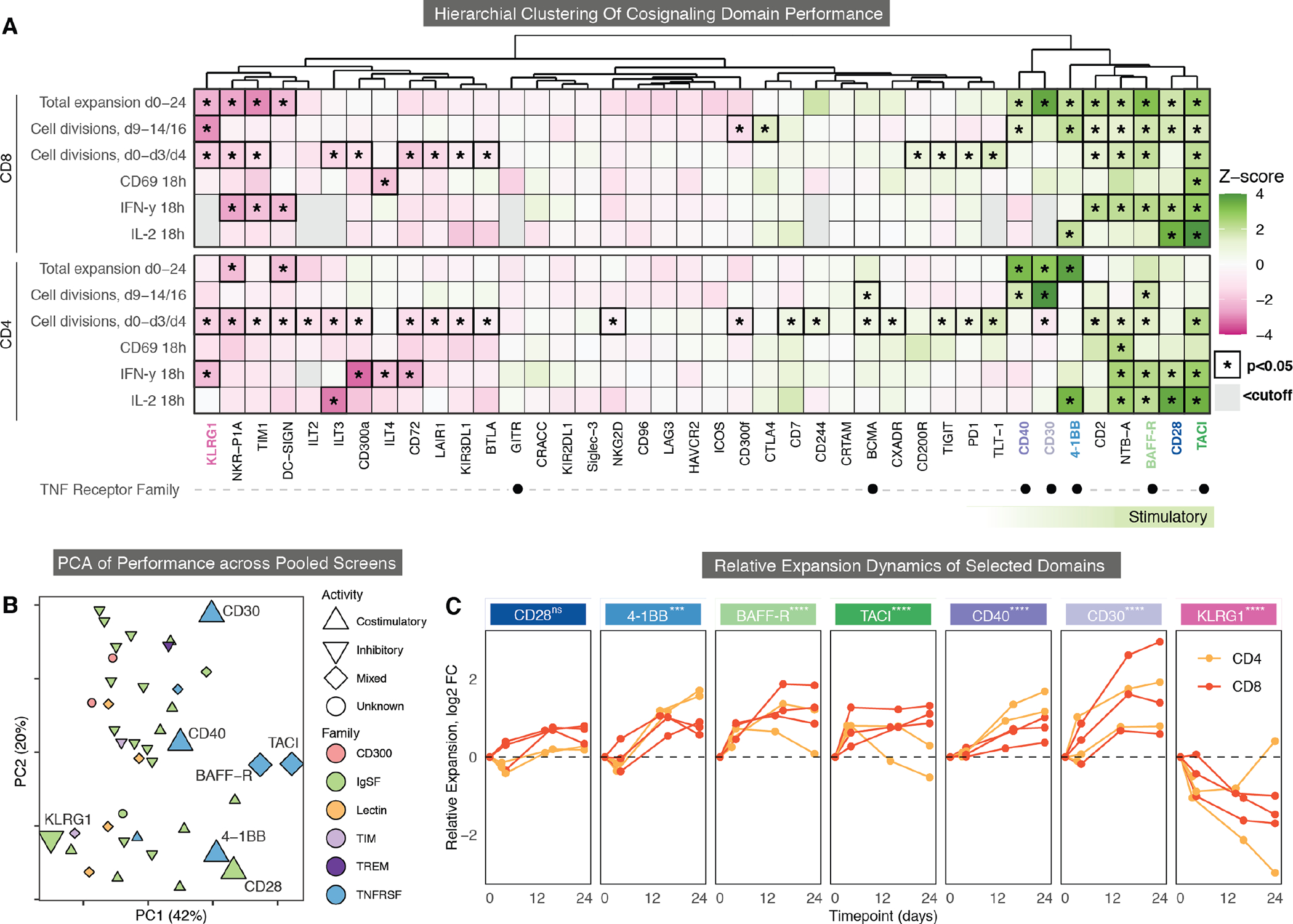
(A) Hierarchical clustering of the CAR signaling domain library in CD4 and CD8 T cells stimulated with CD19+ K562 tumor cells. All 40 domains were clustered based on their z-scores in each assay. 8 stimulatory domains were identified on the right (green bar). Gray boxes are excluded domains where < 500 cells were detected for the assay. TNF receptor family members are marked with black dots. KLRG1 (pink), CD40 (dark purple), CD30 (light purple), 4-1BB (light blue), BAFF-R (light green), CD28 (dark blue), and TACI (dark green) are highlighted. Significance is indicated by a black border and an asterisk(Wilcoxon rank-sum test, FDR < 0.05). (B) Principal components analysis (PCA) of pooled library screen cytokine, proliferation, expansion, and activation data is shown for CD4 T cells, CD8 T cells, CD19+ and CD19− stimulation conditions, and all donors and timepoints. Chosen CARs are larger, with shapes and color indicating known function and protein family. (C) Fold-change of the proportion of selected CARs within the library at each timepoint (x-axis) over 24 days of repeated stimulation with irradiated CD19+ K562 cells as compared to the average CAR in the pooled library. CARs were measured in CD4 and CD8 primary human T cells individually in 2 to 3 biological replicates. Significant p values were derived from a linear mixed effects model; FDR < 0.05:*, < 0.01:**, <0.001:***, <0.0001:****.