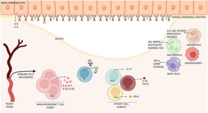
FIGURE 1.
Schematic representation of immune cells and molecules involved in the pathogenesis of bullous pemphigoid. Bullous pemphigoid is determined by IgG, IgE attaching BP180 located in the dermal-epidermal junction. Epidermal cells react by releasing interleukin (IL) 6 and 8. Eventually, this process leads to the recruitment of immune cells (mast cells, macrophages, and eosinophils) which infiltrate the skin and release inflammatory interleukins (IL) and proteolytic enzymes. T cells contribute to this inflammatory process by releasing interleukins at both peripheral (blood) and lesional (skin) level. Especially IL-4, IL-13, IL-31 are crucially involved in B cell proliferation, antibody production and Ig-class switching, itch and eosinophils activation, while IL-17 support neutrophil recruitment. Together, immune cells induce expression of chemokines, thus increasing skin infiltration. The result of this process is the formation of erythematous urticarial plaques and, later, dermal-epidermal splitting causative of blistering. IL, interleukin; Ig, immunoglobulin; Th, T helper; TGF-β, tumor growth factor β; IFN-ɣ, interferon ɣ; GZMB, granzyme B; MMP9, matrix-metallopeptidase 9; LB5, leukotriene B 5; ROS, reactive oxygen species; C5, complement component 5.

