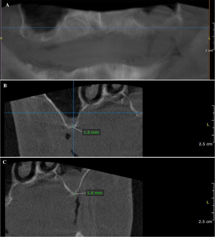
Fig. 1.
Radiographic evaluation of an alveolar ridge defect with a Type-III bone defect in the maxilla before augmentation. A Panoramic radiograph demonstrated massive bone loss in the right and left maxilla. The patient showed local swelling of the basal maxillary sinus mucosa during the preliminary examination. After assessment by an otorhinolaryngologist, the patient had a clinically asymptomatic situation and there was no objection to augmentation of the sinus. B Sagittal section of the CBCT illustrating the region of interest in the first quadrant. C Sagittal section of the CBCT illustrating the region of interest in the second quadrant. The green line corresponds to the vertical height of the bone in the defect area before augmentation

