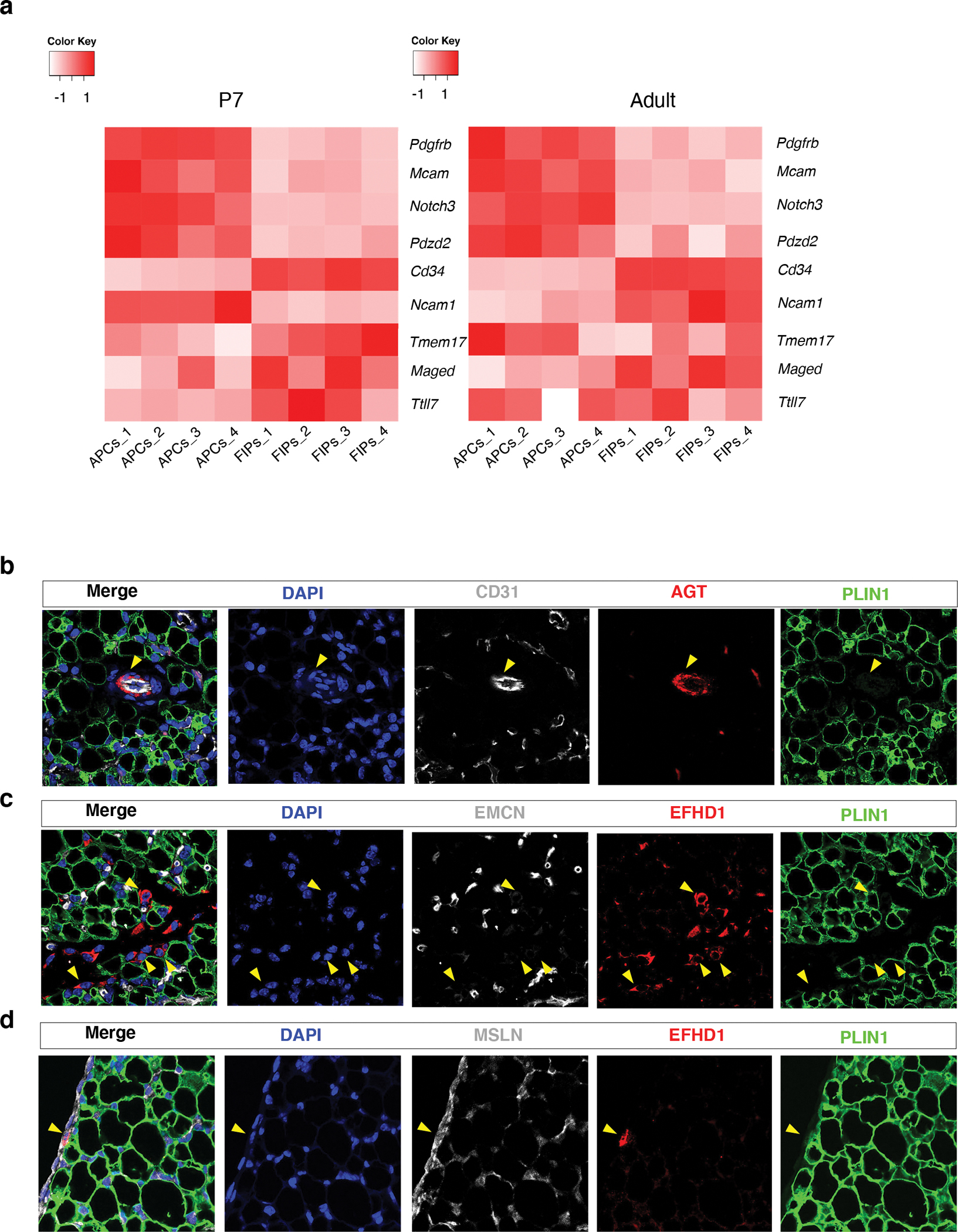
Figure 4. Localization of APCs and FIPs in P7 eWAT.

a) Heatmap depicting the expression of previously reported transcripts 29 defining pericytes vs. perivascular fibroblasts within P7 and adult APCs and FIPs. Enriched expression of Pdgfrb, Mcam, Notch3, and Pdzd2, is characteristic of pericytes, whereas enriched expression of Cd34, Ncam1, Tmem17, Maged, and Ttll7, is more charactertistic of fibroblasts 29. Expression normalized by z-score.
b) Representative 63x magnification confocal image of CD31, ANGIOTENSINOGEN (AGT), and PERILIPIN (PLIN) expression in P7 eWAT sections. Nuclei were counterstained with DAPI. 3 independent experiments were conducted with similar results.
c) Representative 63x magnification confocal image of ENDOMUCIN (EMCN), EFHD1, and PLIN, expression in P7 eWAT sections. Nuclei were counterstained with DAPI. 3 independent experiments were conducted with similar results.
d) Representative 63x magnification confocal image of MESOTHELIN (MSLN), EFHD1, and PLIN, expression in P7 eWAT sections. Nuclei were counterstained with DAPI. 3 independent experiments were conducted with similar results.
