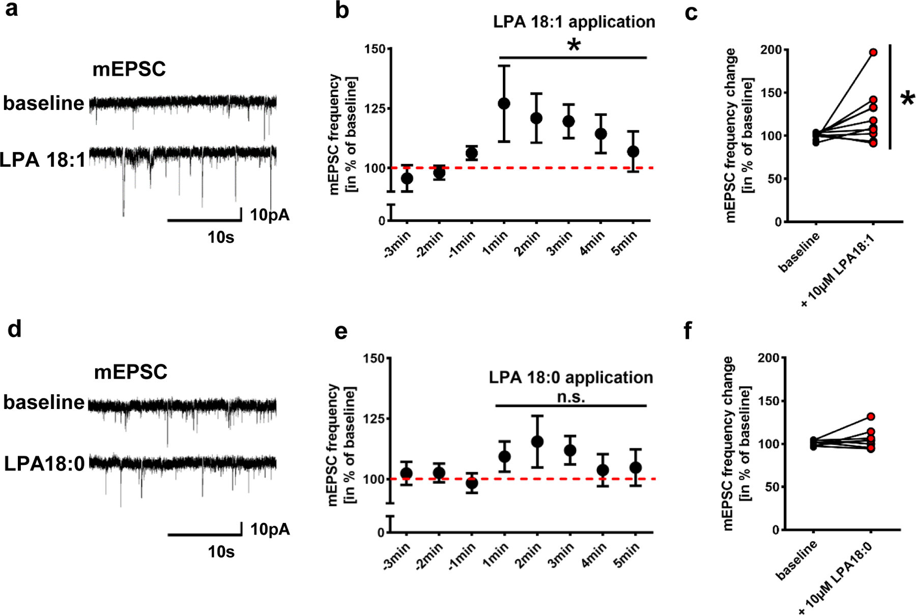
Extended Data Fig. 2. LPA 18:1 is a synaptic active LPA.

a. Original traces of patched neurons before and after LPA 18:1 application suggest higher number of miniature inward currents (mEPSCs) when compared to values before LPA18:1 application
b. Quantitative analysis of mEPSC frequencies following LPA 18:1 application were significantly increased when compared to baseline values before LPA 18:1 application (n = 10; P=0.0327, repeated measures one-way ANOVA). Data was calculated in % of the mean baseline over 5 min before drug application.
c. For better evaluation, mean of the baseline of individual neurons over 3 min before LPA 18:1 stimulation was compared to that during the first 3 min after LPA18:1 application finding significant differences (n = 10, P=0.027, two-tailed Wilcoxon matched-pairs signed rank test).
d. Original traces before and following of LPA 18:0 application show comparable mEPSCs.
e,f. Quantitative analysis revealed no significant effect of LPA 18:0 application on mEPSC frequency (n = 9; repeated measures one-way ANOVA). Before and after LPA 18:0 application mean values of individual neurons were calculated as described above and shown in f (n = 8, two tailed Wilcoxon matched-pairs signed rank test).
Points and bars in b and e represent mean and s.e.m.
