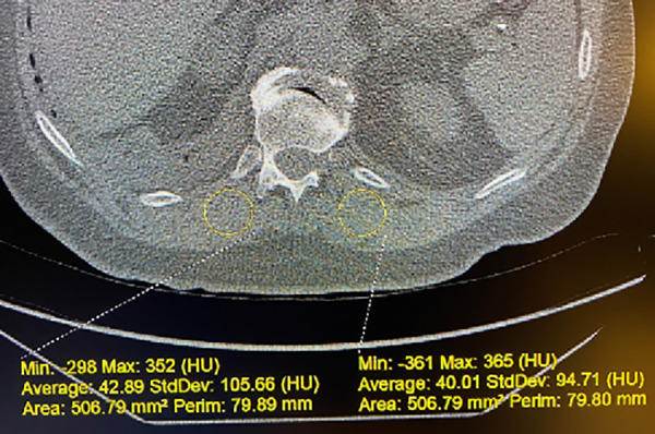
Figure 2.

Example of paraspinous muscle density (PSMD) measurement on noncontrast computed tomography images. The method for measurement of the PSMD at the T12 costovertebral junction is illustrated. The right 12th rib’s junction with the T12 vertebral body is the axial location for the measurement on that scan. A 500-mm2 region of interest is used. In this case, right PSMD has an average value of 42.89 HU, and left PSMD has an average value of 40.01 HU. These two measurements are then averaged to yield the mean PSMD for the participant on this specific examination. HU = Hounsfield units; Max = maximum; Min = minimum; Perim = perimeter.
