Fig. 1.
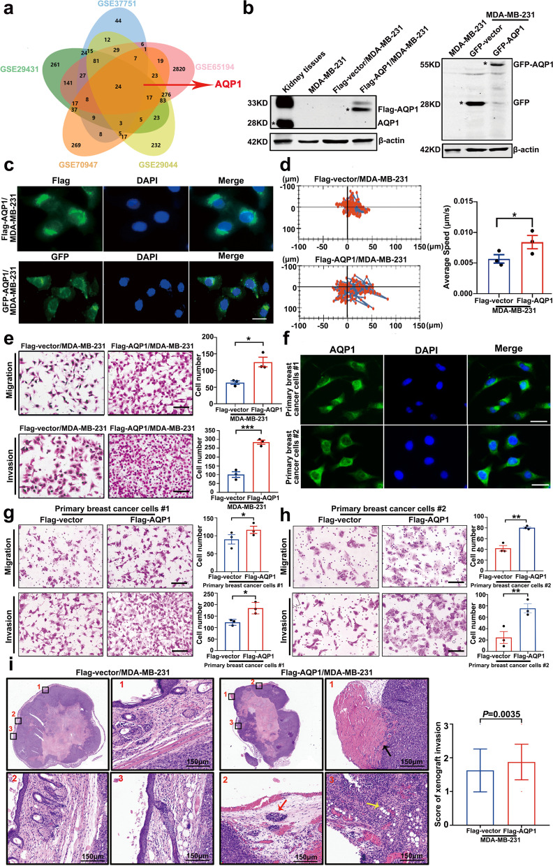
Water channel protein AQP1 mainly localized in the cytoplasm of breast cancer and it was crucial for breast cancer local invasion. a A Venn diagram showed several candidate molecules potentially regulating breast cancer progression based on a combination analysis of five gene expression profiles. b Western blot analysis of AQP1 expression in kidney tissues and MDA-MB-231 cells with two different tags. Kidney tissues were used as a positive control and β-actin was the loading control. c Representative immunofluorescence images of AQP1 localization in MDA-MB-231 cells with two different tags. Scale bar = 20 μm. d Migration tracks of MDA-MB-231 cells stably transfected with control (Flag-vector) or AQP1 (Flag-AQP1) and images were captured every 30 min for 6.5 h. Bar graphs show quantification of cell speed (right). Error bars represent SEM of 3 independent experiments (two-tailed Student’s t test, *P < 0.05). e The abilities of migration and invasion were detected using Flag-vector/MDA-MB-231 and Flag-AQP1/MDA-MB-231 cells. Values were expressed as mean ± SEM from three independent experiments (two-tailed Student’s t test, *P < 0.05, ***P < 0.001). Scale bar = 100 μm. f Immunofluorescence images showed that AQP1 localized in cytoplasm of two primary breast cancer cells. Scale bar = 20 μm. g, h Representative migration or invasion images of control group and AQP1-overexpressing primary breast cancer cells. Values were expressed as mean ± SEM from three independent experiments (two-tailed Student’s t test, *P < 0.05, **P < 0.01). Scale bar = 100 μm. i Representative images of hematoxylin–eosin staining in xenograft paraffin specimens, part of square 1/2/3 was enlarged in picture 1/2/3 respectively (muscle involvement: black arrow, Satellite nodules: red arrow, adipocytes involvement: yellow arrow). N = 12/group. The score of xenografts invasion in mice was analyzed quantitatively. All in vitro experiments were repeated at least 3 times

