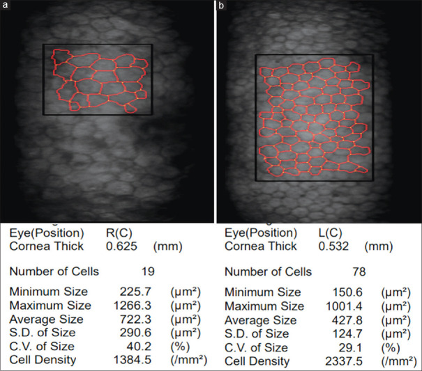
Figure 5.
Specular microscopy images of the AC glass FB. (a) Specular microscopy image of the right eye of the patient in case-1 with an AC glass FB showing pleomorphism and polymegathism with decreased cell density suggestive of endothelial cell loss and decrease in cell count; increase in minimum, maximum, and average cell size; increase in corneal thickness suggestive of endothelial compromise. (b) Specular microscopy image of the normal left eye of the patient in case-1 showing normal cell size, shape, and density for age

