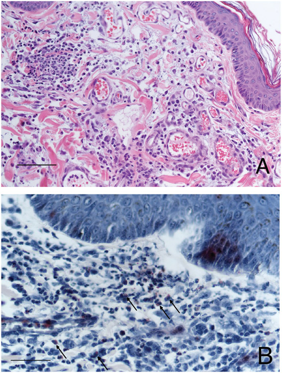
Figure 2—
Photomicrographs of skin biopsy specimens from the dog in Figure 1. A—Note the dermal edema, vascular ectasia, and perivascular to interstitial inflammatory infiltration composed predominantly of eosinophils and neutrophils with fewer macrophages, lymphocytes, and plasma cells. H&E stain; bar = 100 μm. B—Eosinophils (arrows) are highlighted with reddish staining; Luna stain; bar = 50 μm.

