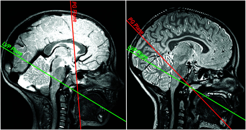
Figure 3.
The midsagittal image is shown for two participants with variations in the orientation of the palatoglossus (PG) muscle. The palatoglossus (PG) muscle plane is in red. The levator veli palatini (LVP) muscle plane is in green. Variations in the PG–LVP angle, primarily due to PG orientation, are visually apparent.

