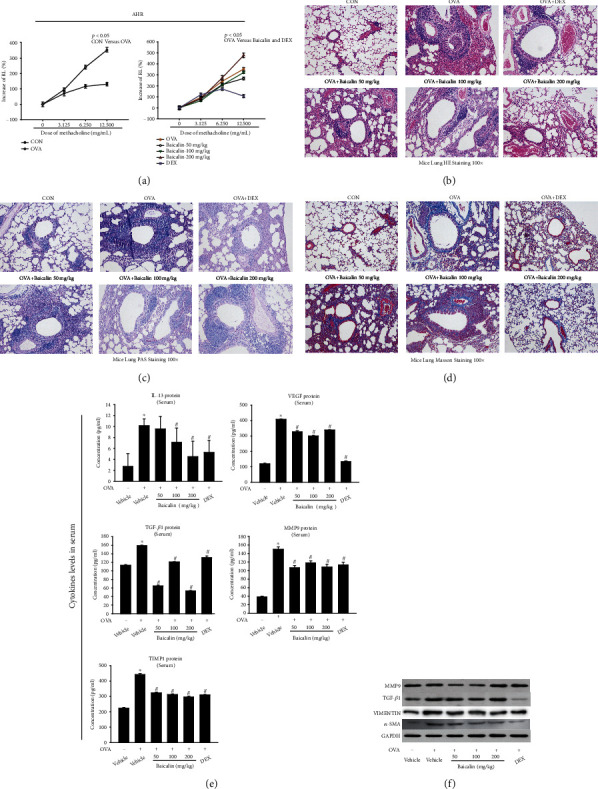
Figure 1.

Effects of baicalin on AHR, inflammatory cell infiltration, lung histology, and airway remodeling. (a) RL was measured after treatment with methacholine. (b) HE staining was used to measure airway wall thickness, bronchial lumen, mucosal edema, and inflammatory cell infiltration. (c) PAS staining was used to measure mucus production. (d) Masson staining was used to measure collagen deposition around the airways (×100 magnification). (e) Protein expression of IL-13, VEGF, TGF-β1, MMP9, and TIMP1in serum was measured by ELISA. (f) Protein expression of α-SMA, VIMENTIN, MMP9, and TGF-β1 in lung tissue was measured by western blot. All data were shown as mean ± SEM. Sections are representative of n ≥ 6 mice from each group. ∗P < 0.05 versus vehicle. #P < 0.05 versus OVA.
