
Figure 2.
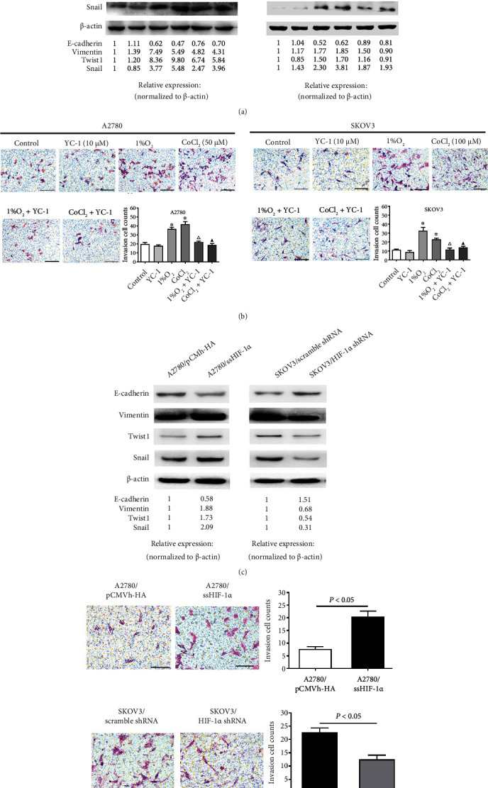
Hypoxia-induced HIF-1α regulates the levels of EMT markers and invasion in EOC cell lines. (a) Relative protein levels of EMT markers in A2780 and SKOV3 after YC-1 treatment under normoxic (21% O2) or hypoxic (1% O2 or CoCl2) conditions, respectively. (b) The invasion ability of A2780 and SKOV3 treated as described for (a) was detected by Matrigel invasion assay. Scale bars: 100 μm. In each experiment, the numbers of cells that penetrated the membrane were counted in five microscopic fields per filter. The invasion cell counts are presented as the mean ± SD of three independent experiments. ∗P < 0.05 compared with the control group, △P < 0.05 compared with the 1% O2 group, and ▲P < 0.05 comparedwith the CoCl2 group by one-way ANOVA with LSD multiple comparison test. (c) Protein levels of EMT markers in transfected A2780 cells overexpressing HIF-1α and transfected SKOV3 cells with repression of HIF-1α under normoxic conditions. (d) The invasion ability of transfected A2780 cells overexpressing HIF-1α and transfected SKOV3 cells with repression of HIF-1α was detected by Matrigel invasion assay. Scale bars: 100 μm.