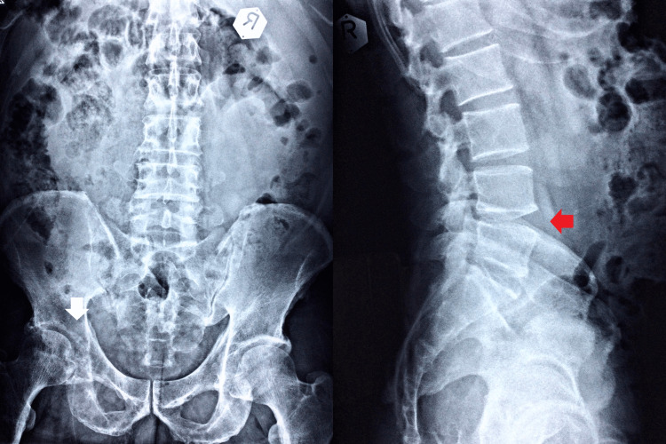
Figure 1. Lumbar and pelvic radiography.
The anteroposterior and lateral views appear to have reduced L4-5 disc space and a marginal anterior osteophyte in the lumbosacral spine (LS) vertebral body (red arrow). Multiple lucent foci are also found on the left pelvic rim, femoral head, and proximal body (white arrow).

