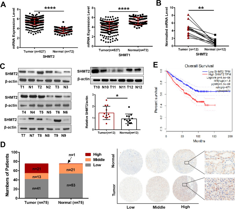
Fig. 1. SHMT2 was aberrantly upregulated in ccRCC tissues and associated with patient survival.
A Relative expression of SHMT2 and SHMT1 in ccRCC specimens and controls based on the TCGA database. B qRT-PCR analysis was performed to evaluate the SHMT2 mRNA levels in 12 pairs of ccRCC tissues and controls. C Western blot analysis was performed to evaluate the SHMT2 protein levels in 12 pairs of ccRCC tissues and controls. β-actin expression was detected as a loading control. T meaned tumors and N meaned paired controls. D TMA IHC scores showing that high, middle and low SHMT2 expression were observed in 21, 13, and 41 of the 75 tumor specimens. The corresponding numbers were 1, 21, and 53 in the 75 paired control tissues (left). Representative IHC staining of SHMT2 protein in human ccRCC tissues (right). E Overall survival curves of ccRCC patients with different SHMT2 expression levels based on the GEPIA database. *P < 0.05, **P < 0.01, ****P < 0.0001.

