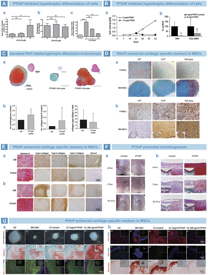
FIGURE 7.
PTH-related peptide (PTHrP) in osteoarthritis. (A). PTHrP prevented the hypertrophy of chondrocytes. Collagen quantification results that the content ratio of collagen type II to collagen type I was significantly improved by PTHrP (A), while the proteoglycan exhibited no difference by DMMB assay (B). However, the expression of collagen type X was significantly downregulated by PTHrP, as qPCR results (Kafienah et al., 2007). (B). PTHrP inhibited the endochondral indexes of ALP activity and mRNA expression of Indian hedgehog and collagen type X (Fischer et al., 2010). (C). Pulsative stimulation of PTHrP was performed on chondrocytes (A). The deposition of proteoglycan and collagen type II was promoted (B), and decreased expression trend of collagen type X was found (C) (Fischer et al., 2014). (D). Supplemented PTHrP from fourth day strengthened the cartilage matrix deposition of periosteal MSCs and bone marrow MSCs by Alcian blue (A), but suppressed the endochondral osteogenesis, as immunohistochemical (IHC) staining of collagen type X (B) (Rajagopal et al., 2021). (E). PTHrP improved the chondrogenic matrix deposition of proteoglycan and collagen type II by Safranin-O staining and IHC staining (A). However, the markers of endochondral osteogenesis were inhibited, such as collagen type I, collagen type X and Runx-2 by IHC staining (Kim et al., 2008). (F). The time window between 4 and 6 weeks for PTHrP injection benefited the rat knee cartilage repair best, as the result of gross morphology (A) and Safranin-O staining (B) (Zhang et al., 2013). (G). Implanted cell pellets that treated with PTHrP showed improved Safranin-O staining and anti-collagen type I/II IF staining after 3 weeks (A). Meanwhile, weakly positive stains of Alizarin Red S and anti-collagen type X/CD31 IF staining were found (B) (Anderson-Baron et al., 2021).

