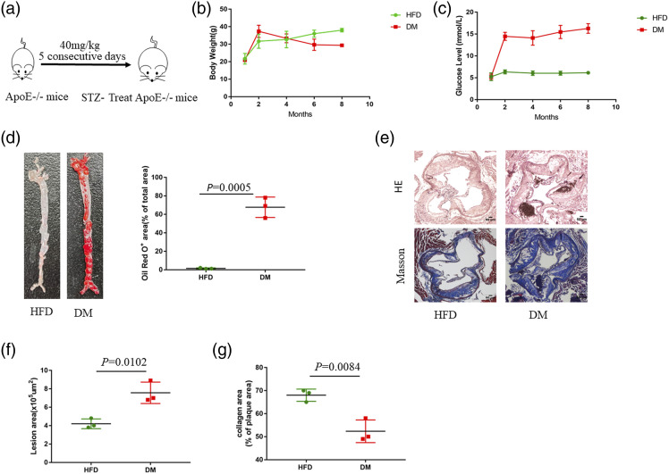
Figure 1.
Diabetes accelerates the development of atherosclerosis in ApoE−/− mice. (a) ApoE−/− mice were fed an HFD diet and treated with STZ for 5 days to establish a diabetic atherosclerotic model. (b) Body weight (g) (n = 3), (c) Blood glucose (mmol/L) (n = 3). Results are expressed as mean ± SEM (d) Oil Red O staining of aortas from ApoE−/− mice fed an HFD for 24 weeks (n = 3). Data represent the percentage of plaque area/total vessel area. Unpaired two-tailed Student’s t-test (n = 3). (e) H&E staining (the first row) and Masson staining (the second row) of representative aortic root sections (n = 3). Scale bar, 50 μm. (f) Quantitative analysis of the lesion area. Unpaired two-tailed Student’s t-test. (g) Quantitative analysis of plaque collagen area. Unpaired two-tailed Student’s t-test.

