Fig. 1.
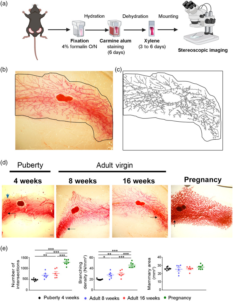
Imaging and semiquantification of mammary gland epithelium stained with carmine alum. (a) Schematic overview of the procedure followed for carmine alum staining of the mammary glands. Created with BioRender.com. (b) Representative whole mount carmine alum staining and (c) its skeletonized image that was used to perform Sholl analysis in an abdominal mammary gland from an 8-week-old virgin C57BL/6 mouse. Black outline defines the boundaries of the mammary fat pad. (d) Representative images of whole mounts of abdominal mammary glands stained with carmine alum at different stages (4, 8, and 16 weeks and pregnancy at 15.5 dpc). The high background is shown with black arrows, whereas the rudimentary duct at puberty by blue arrow. Scale bar = 1 mm, magnification = 2×. (e) Quantitative analysis of intersection number, branching density and mammary fat pad area at the distinct developmental stages through Sholl analysis ( to 7 per group). Data are shown as mean values ± SEM, one-way ANOVA and Tukey post-hoc test was performed for statistical analysis (*, **, ***).

