Fig. 4.
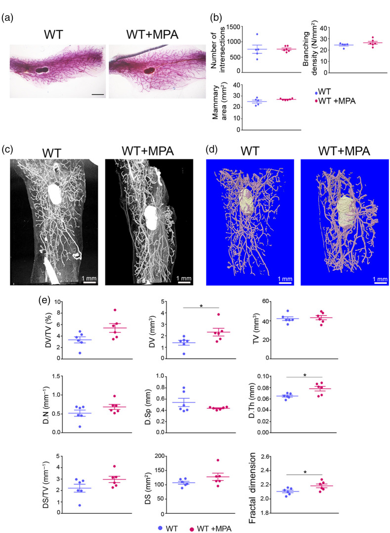
Effect of short-term administration of MPA in the mammary glands. (a) Representative whole mount carmine alum staining images of abdominal mammary glands from MPA-treated and WT control female mice. Scale bar = 1 mm, magnification = 4×. (b) Semiquantitative analysis of intersection number, branching density and total mammary gland area using carmine alum staining, and Sholl analysis ( to 6 per group). (c) Representative 2D and (d) 3D reconstructed images through PTA-enhanced microCT of the mammary epithelium from MPA-treated and WT control mice. (e) Comparative quantitative analysis of mammary gland epithelium between MPA-treated and WT control mice employing PTA-enhanced microCT (). Data are shown as mean values ± SEM. Unpaired -test was performed for statistical analysis (*).

