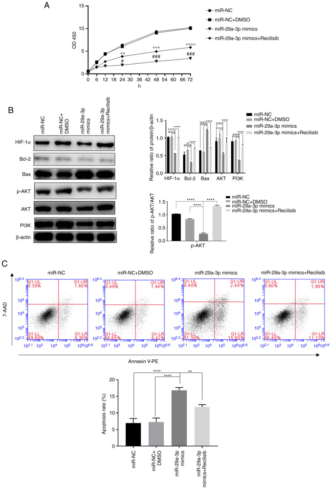
Figure 4.
The effect of miR-29a-3p via the PI3K/AKT/HIF-1α pathway on glioma cells' proliferation and apoptosis levels. (A) The proliferation level was detected by CCK-8 assay, **P<0.01, ***P<0.001, ****P<0.0001 vs. miR-NC + DMSO; #P<0.05, ###P<0.001 vs. miR-NC. (B) The expression levels of HIF-1α, Bcl-2, Bax, AKT, PI3K and the ratio of p-AKT/AKT, were detected by western blotting, **P<0.01, ****P<0.0001. (C) The levels of apoptosis in each group were detected by flow cytometry, **P<0.01, ****P<0.0001. All data were expressed as the mean ± standard deviation (n=3). miR, microRNA; HIF-1α, hypoxia-inducible factor 1; NC, negative control; HIF-1α, hypoxia-inducible factor 1; p-, phosphorylated; PE, phycoerythrin.

