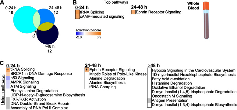
Fig. 4.
Enriched pathways in whole blood. A Venn diagram represents all enriched pathways at each of the time points (0–24 h, 24–48 h, >48 h; Fisher’s p-value<0.05). There was no overlap of pathways between the time points. B Top over-represented pathways with significant z-score (z ≥ 2, predicted activation) at 0–24 h and 24–48 h. No significant z-scores were predicted for pathways at >48 h. C Top over-represented pathways unique to each TP (Fisher’s p-value<0.05). Orange cells indicate positive z-score for the pathway, and blue indicate negative z-score. White bars indicate no direction can be predicted, and grey bars indicate no prediction can be performed. Up arrows indicate predicted significant activation (z ≥ 2)

