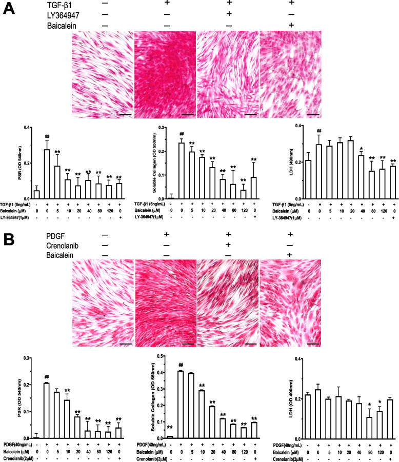
Fig. 1.
Baicalein inhibits TGF-β1- (A) or PDGF (B)-induced fibrosis in CCC-ESF-1 cells. Cells were seeded in collagen I-coated plates and treated/not treated with TGFβ1 (5 ng/mL) or PDGF (40 ng/mL) in the presence or absence of baicalein (5–120 μM) for 48 h. Total collagen deposition was visualized by PSR staining and quantified by spectrophotometry. Soluble collagen in the supernatant was detected by the Sircol Soluble Collagen Assay. Cytotoxicity was assessed using the lactate dehydrogenase release (LDH) assay. Representative images were shown, scale bar = 200 μm. Quantitative data were the mean ± SD from one representative experiment out of ≥ 3 experiments with similar results, n ≥ 4 wells per group. #P < 0.05, ##P < 0.01 vs. no TGF-β1 or PDGF group; *P < 0.05, **P < 0.01 vs. TGF-β1- or PDGF-only group

