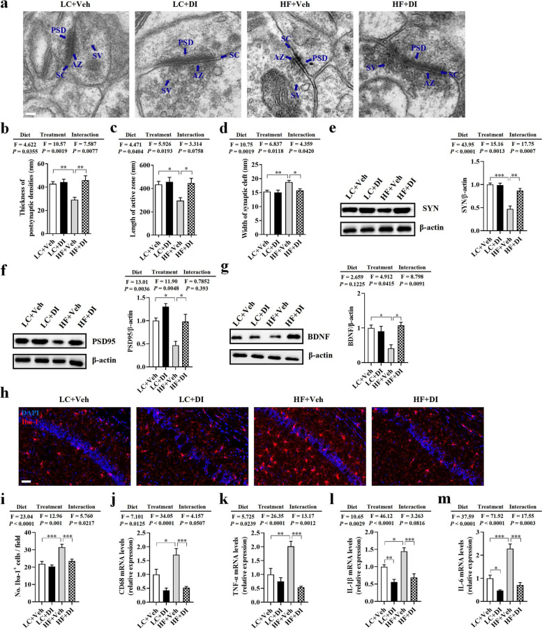
Fig. 3.
DI supplementation mitigated synaptic impairment and neuroinflammation in the hippocampus of mice on HF diet. a Representative ultrastructure of synapses in the cornu ammonis 1 (CA1) region of mice on the electron micrograph (scale bar: 100 nm). b–d Image analysis of the thickness of postsynaptic density (PSD), length of the active zone (AZ), and width of the synaptic cleft (SC) (n = 2, 8 images per mouse). e–g The protein expression levels of SYN, PSD95, and BDNF in the hippocampus (n = 5). h The immunofluorescent staining of Iba-1 in CA1 of the hippocampus. i The quantification of Iba-1+ cells in CA1 of the hippocampus (n = 3, 5 images per mouse, scale bar: 50 μm). j–m The mRNA expression of CD68, TNF-α, IL-1β, and IL-6 in the hippocampus (n = 6). Values are mean ± SEM. *P < 0.05, **P < 0.01, ***P < 0.001

