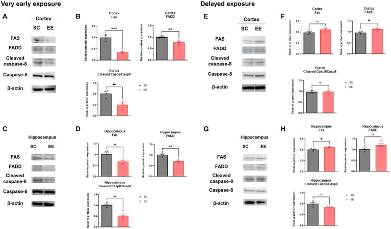
Figure 7.
Effects of very early and delayed exposure to EE on protein expression in the extrinsic apoptosis pathway in the cerebral cortex and the hippocampus. (A,C) Representative WB images from cerebral cortex (A) and hippocampus (C) obtained after very early EE exposure. (B,D) Quantification of Fas, FADD, and cleaved Caspase-8 levels in the cerebral cortex (B) and the hippocampus (D) (n = 4 per group). (E,G) Representative WB images from cortical (E) and hippocampus (G) obtained after delayed EE exposure. (F,H) Quantification of Fas, FADD, and cleaved Caspase-8 in the cerebral cortex (F) and hippocampus (H) (n = 4 per group). All statistical comparisons were performed via Student’s t-tests. Data represented are means ± SEM. *p < 0.05, **p < 0.01, and ***p < 0.001. EE, environmental enrichment; WB, western blot.

