Fig. 1.
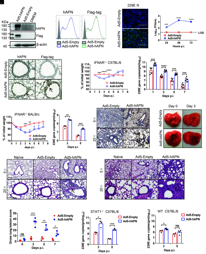
Development of mice sensitized to 229E infection. (A and B) To assess hAPN expression and surface localization, 17CL-1 cells were transduced with Ad5-hAPN or Ad5-Empty with MOI of 20 at 37 °C for 4 h. The hAPN expression was monitored by western blot assay (A) or flow cytometry (B). (C and D) Ad5-hAPN-transduced 17CL-1 cells were infected with 229E at MOI of 0.04 at 48 h post transduction, and viral antigen was determined by immunofluorescence assays at 48 h.p.i. (C), and viral titers were determined by focus forming assay (FFA) at 24, 48, and 72 h.p.i. (D). (Scale bars = 200 µm.) (E) Five days after transduction with 2.5 × 10⁸ FFU of Ad5-hAPN or Ad5-Empty in 75 μL DMEM intranasally, lungs were harvested from mice, fixed in zinc formalin, and embedded in paraffin. Sections were stained with an anti-hAPN antibody and anti-Flag antibody (brown color). Arrowheads indicate regions with hAPN expression. (Scale bars = 100 µm.) (F and G) Ad5-hAPN or Ad5-Empty-transduced IFNAR−/− C57BL/6 and BALB/c mice were intranasally infected with 1.5 × 10⁵ TCID50 of 229E in 75 μL DMEM. Weight changes in 6 to 8-wk-old IFNAR−/− C57BL/6 (F) and BALB/c mice (G) were monitored daily (n ≥ 3 mice per group). To obtain viral replication kinetics in IFNAR−/− C57BL/6 (F) and BALB/c mice (G), lungs were harvested and homogenized at the indicated time points, and the viral load was detected by RT-qPCR. Viral loads are expressed as gene copies /g lung tissue (n = 3 mice per group per time point). Data are representative of two independent experiments. (H) Three days post infection, lungs were harvested from IFNAR−/− C57BL/6 mice, fixed in zinc formalin, and embedded in paraffin. Sections were stained with a rabbit anti-229E nucleocapsid protein polyclonal antibody. Arrowheads indicate regions with 229E antigen expression. (Bars = 200 and 50 μm, Top and Bottom, respectively). (I and J) Representative HE staining of lungs from IFNAR−/− C57BL/6(I) and BALB/c (J) mice harvested at 3 d.p.i. Arrowheads indicate regions with interstitial pneumonia with perivascular and interstitial inflammatory cell infiltrates. (Bars = 200 and 100 μm, Top and Bottom, respectively). (K) Photographs of lung specimens isolated from infected IFNAR−/− C57BL/6 mice at indicated time points are shown. Arrowheads indicate regions with vascular congestion and hemorrhage. (L) Gross lung lesion scores are mean ± SE (error bars) and were graded based on the percentage of lung area affected (n = 3 or 4 mice per group per time point). (M and N) Ad5-hAPN or Ad5-Empty-transduced STAT1−/− C57BL/6(L) and WT C57BL/6 (M) mice were intranasally infected with 1.5 × 10⁵ TCID50 of 229E in 75 μL DMEM. Lungs were harvested and homogenized at the indicated time points, and the viral load was detected by RT-qPCR. Viral loads are expressed as gene copies /g lung tissue (n = 3 mice per group per time point). Data are representative of two independent experiments. (*P ≤ 0.05, **P ≤ 0.005, ***P ≤ 0.0005, ****P ≤ 0.0001).

