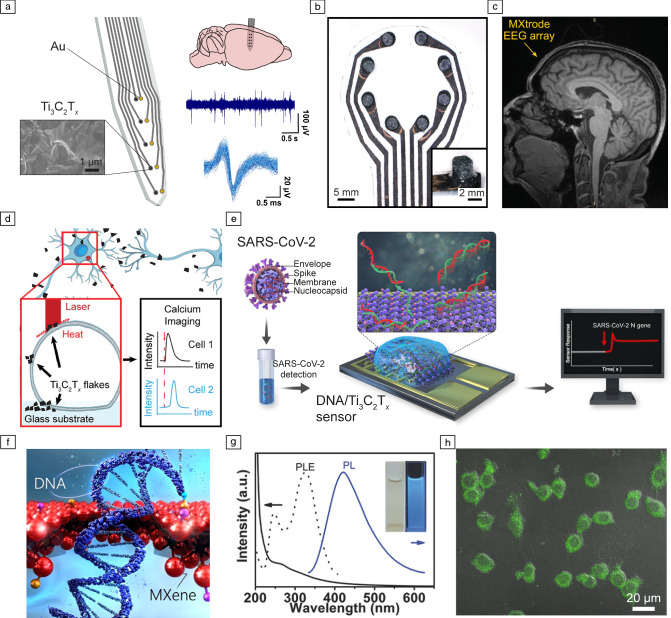
Figure 2.
Bioelectronics and multimodal biosensing with MXenes. (a) Schematics of an intracortical array patterned with 25 µm Ti3C2Tx and adjacent Au contacts for recording neural activity in vivo. The close-up view shows a scanning electron microscopy image of the surface of one Ti3C2Tx contact. The arrays can record in vivo intracortical neural activity with higher signal-to-noise ratio and reduced susceptibility to 60 Hz interference than Au (reproduced with permission from Reference 21). (b) Photograph of Ti3C2Tx-infused electrode arrays for electroencephalography (EEG). (c) MXene EEG electrode array placed on human forehead and imaged in 3T clinical MRI scanner using T1-weighted magnetization prepared rapid acquisition gradient echo. (b, c) Reproduced with permission from Reference 23. (d) Schematics of the photothermal stimulation of a dorsal root ganglion (DRG) neuron network with Ti3C2Tx flakes (reproduced with permission from Reference 30). (e) Schematics illustrating the operation of ssDNA/Ti3C2Tx sensors for the detection of SARS-CoV-2 nucleocapsid (N) gene (reproduced with permission from Reference 39). (f) Schematic illustrating DNA translocation through a MXene nanopore sensor (reproduced with permission from Reference 44). (g) Photoluminescent Ti3C2Tx MXene quantum dots (MQDs) for multicolor cellular imaging. UV–vis spectra (solid line), photoluminescence excitation (PLE) (dashed line), and photoluminescence (PL) spectra (solid line, Ex = 320 nm) of MQD-100 in aqueous solutions. (h) Merged bright-field and confocal images (Ex = 488 nm) of MQD-100 interfaced with RAW 264.7 cells. (g, h) Reproduced with permission from Reference 48.

