FIG. 3.

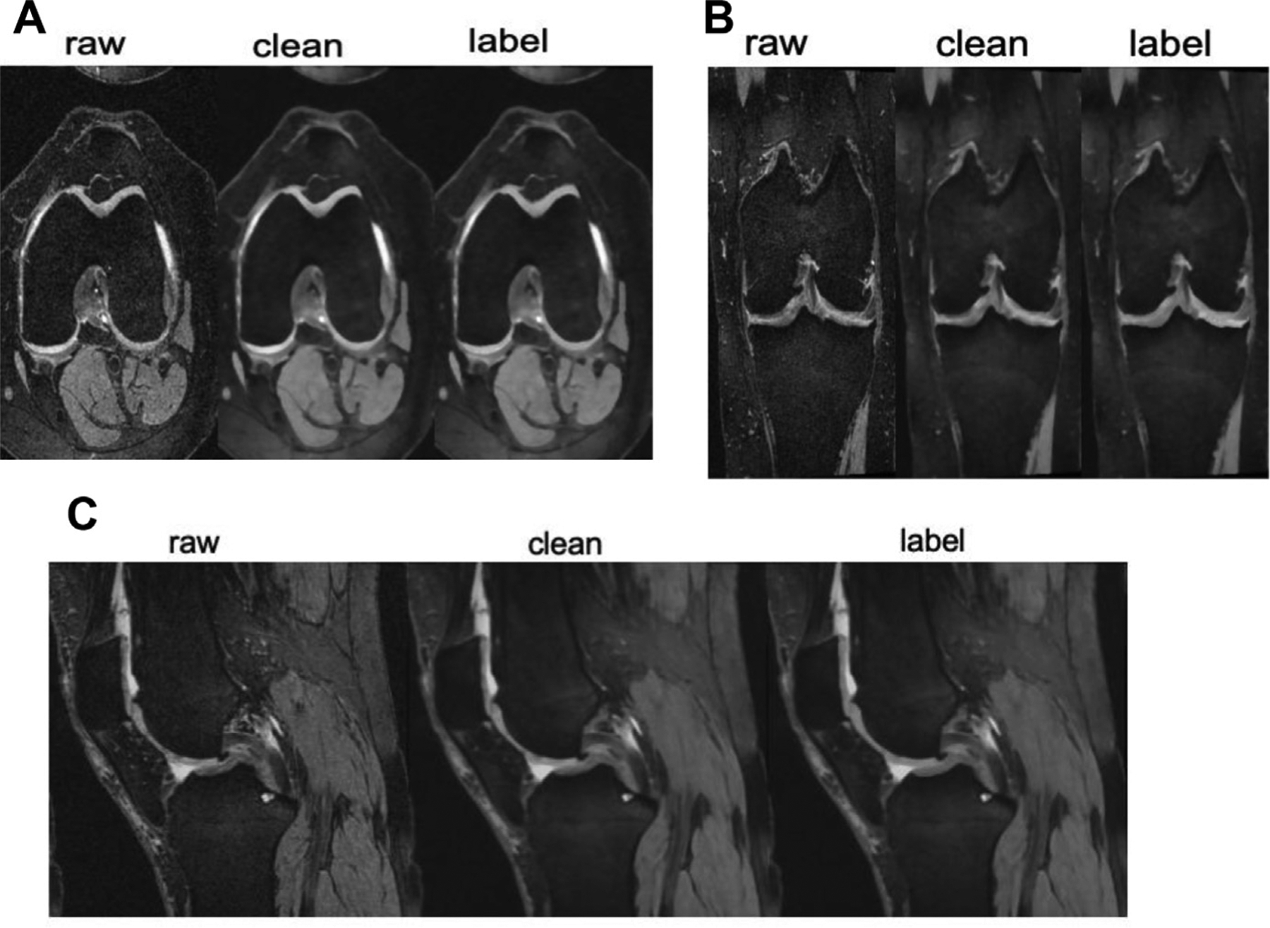
Axial (A), coronal (B), and sagittal (C) knee MRI. Raw demonstrates the images before preprocessing. Clean demonstrates the images after preprocessing. Tissue coloring for segmentation is referred to as the “labeled” image.

Axial (A), coronal (B), and sagittal (C) knee MRI. Raw demonstrates the images before preprocessing. Clean demonstrates the images after preprocessing. Tissue coloring for segmentation is referred to as the “labeled” image.