. 2023 Feb 8;12:e78689. doi: 10.7554/eLife.78689

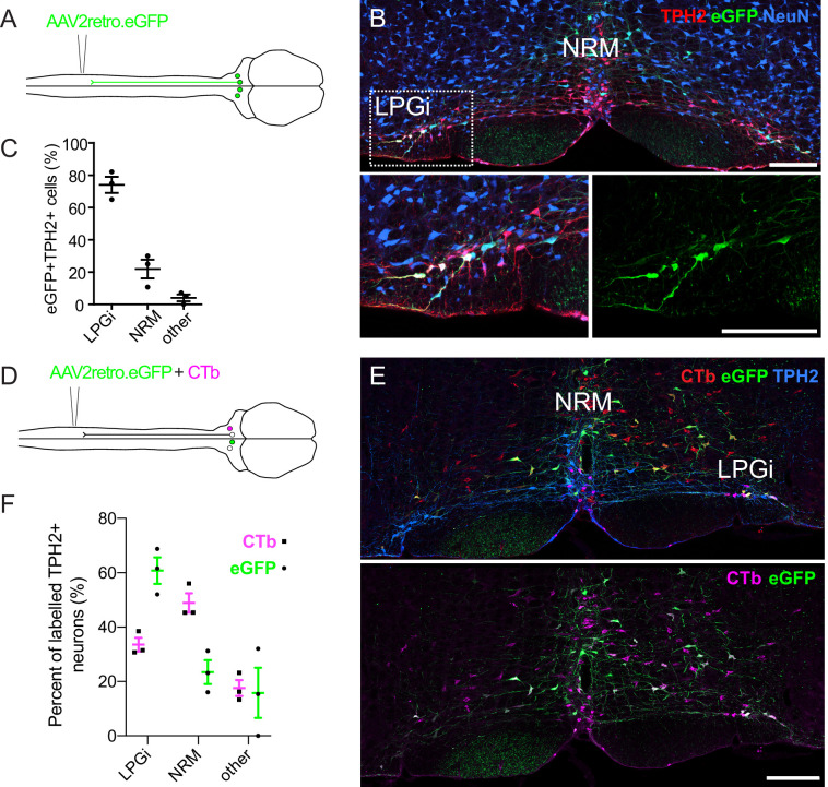
Figure 1. Retrograde labeling of spinally projecting serotonergic neurons with AAV2retro vectors and Cholera toxin b subunit.

(A). Injection scheme for retrograde labeling of spinally projecting neurons with AAV2retro.eGFP. (B) Image of the ventral hindbrain containing eGFP-labeled neurons (scale bar = 200 μm). Inset shows enlargement of the LPGi to reveal eGFP neurons that also express TPH2 (scale bar = 200 μm). (C). Quantification of cell location for eGFP-labeled neurons that express TPH2, each datapoint is a count per animal (n=3) (D). Injection scheme for retrograde tracing from the spinal dorsal horn with AAV2retro.eGFP and CTb. (E). Representative image of the ventral hindbrain containing CTb-labeled and AAV2retro-transduced projection neurons (scale bar = 200 μm). (F). Anatomical locations of retrogradely labeled TPH2 +hindbrain neurons labeled with CTb or AAV2retro (n=3 animals).

Figure 1.

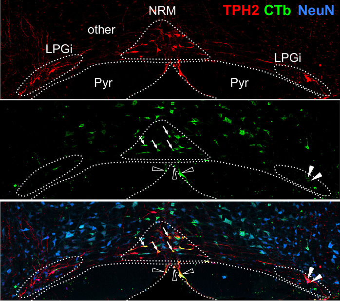
Figure 1—figure supplement 1. Defined areas in the RVM used for quantifying the location of retrogradely labeled neurons.

Figure 1—figure supplement 1.

The NRM was identified as a dense cluster of TPH2-expressing neurons around the midline in a triangular shape, whereas the LPGi were distinguished by their lateral location and separation from the midline structures. All other regions were defined as ‘other’. Examples of retrogradely labeled TPH2 +neurons in the NRM are indicated with arrows, the LPGi with filled arrowheads, and other with empty arrowheads, NRM = nucleus raphe magnus, LPGi = lateral paragigantocellularis, Pyr = Pyramids, (scale bar = 200 μm).
Figure 1—figure supplement 2. Retrograde labeling of serotonergic hindbrain neurons with CTb.

Figure 1—figure supplement 2.

(A). injection scheme for CTb tracing from the lumbar dorsal horn and an example of the injection site in the lumbar spinal cord (scale bar = 200 μm). (B). Example of a hindbrain section from the injection site shown in A (scale bar = 200 μm). The inset shows that many CTb-labeled neurons that express TPH2 are found in the nucleus raphe magnus (indicated by arrows). (C). Location of CTb-labeled cells in the RVM that express TPH2, each datapoint is a count per animal (n=2).
Figure 1—figure supplement 3. Proportion of neurons retrogradely labeled with CTb and AAV2retro in different RVM areas.
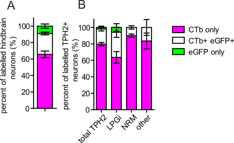
Figure 1—figure supplement 3.

(A). Injection scheme for labeling descending projection neurons with AAV2retro and CTb. (B). Quantification of coexpression of CTb and eGFP in all retrogradely labeled RVM neurons, and (C). Quantification of coexpression of CTb and eGFP in retrogradely traced RVM neurons that contain TPH2. The proportion of retrogradely cells in each area expressing CTb, eGFP, or both eGFP and CTb are illustrated. The cell counts used to generate these charts are given in Table 1 (n=3 animals).
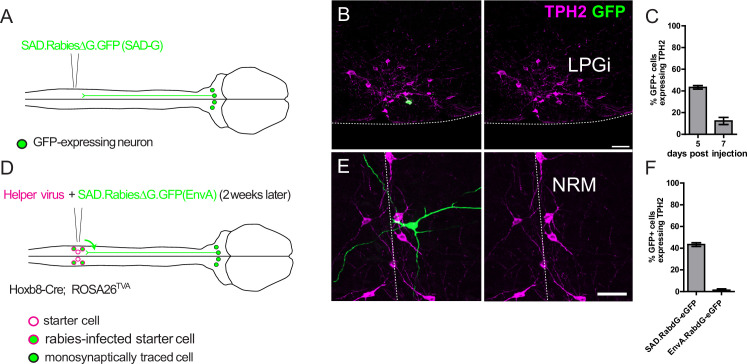
Figure 1—figure supplement 4. labeling of spinally projecting serotonergic neurons with direct rabies infection and transsynaptic rabies tracing.

Figure 1—figure supplement 4.

(A). Injection strategy for the direct labeling of spinally projecting hindbrain neurons with a SAD pseudotyped and G-protein deficient rabies virus. (B). Example of a cell body of a labeled hindbrain neuron that is immunoreactive for TPH2 (scale bar = 50 μm), the ventral border of the hindbrain is indicated by the dotted line. (C). Quantification of the GFP-labeled hindbrain neurons that express detectable levels of TPH2. Note that 7 days after rabies virus injection there are fewer labeled neurons that express TPH2. N=3 animals. (D). Injection strategy for the transsynaptic tracing of neurons from spinal dorsal horn neurons. Hoxb8-Cre is transiently expressed in all lumbar dorsal horn neurons and is crossed with ROSA26TVA to induce TVA expression in all spinal neurons. E. Example of an RVM neuron traced transsynaptically from the spinal dorsal horn (scale bar = 50 μm) (F). Quantification of GFP-labeled neurons in the hindbrain that express detectable TPH2. Data from C. ‘5 days post injection’ is included to allow comparison between directly labeled and transsynaptically traced neurons. N=4 animals for transsynaptic tracing experiment.