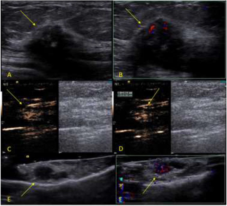
Figure 3:
Example of a malignant study case. The subject is a 67 years-old female patient diagnosed with an invasive ductal carcinoma breast cancer located on the left breast at 2 o’clock position measuring 2.1 cm. After the surgical excision, the SLN was sent to pathology, which determine to be positive for metastatic disease. The SLN was positive for the presence of blue dye, radioactive tracer and UCA at the time of the excision. A B-mode image of the tumor (arrow). B, Color Doppler image of the tumor (arrow). C, Dual-image CEUS and B-mode of the SLN (arrow). D, Dual-image CEUS and B-mode of the SLN with measurement (arrow). E, B-mode image of the ex-vivo specimen of the SLN seen in C and D (arrow). F, Color Doppler image of the ex-vivo specimen of the SLN seen in C and D showing the uptake of UCA (arrow).

