Figure 1.
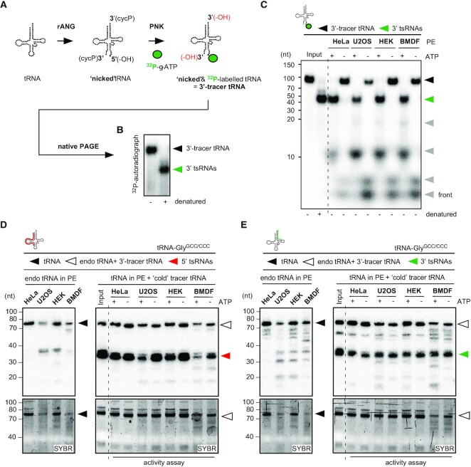
Cytoplasmic protein extracts contain activities which separate and degrade 3‘-tracer tRNAs. (A) Schematic representation of enzymatic reactions for the production of 3′-tracer tRNAs. 32P-γ-ATP-labelling of tRNAs that were hydrolysed by recombinant ANG. Position of the label (32P) on 3' tsRNAs is marked as a green dot. rANG, recombinant Angiogenin; PNK, T4 polynucleotide kinase; cycP, 2′-3′ cyclic phosphate; -OH in black, hydroxyl moiety as result of rANG activity; -OH in red, hydroxyl moieties as result of PNK activity. (B) Representative nPAGE of 32P-γ-ATP-labelled 3′-tracer tRNAs before and after heat denaturation. Black arrowhead, duplex of 3′-tracer tRNAs; green arrowhead, 3′ tsRNAs. (C) 3′-tracer tRNAs (8 nM final) were incubated with cytoplasmic protein extracts (2 μg) obtained from different cell lines in the presence or absence of 2 mM ATP. Reactions were separated by nPAGE and 3′ tsRNA signals were collected by exposing PAA gels to phosphor-imaging plates for ≤ 2 h. Black arrowhead, 3′-tracer tRNAs; green arrowhead, 3′ tsRNAs; grey arrowheads, signals from 3′ tsRNA degradation; PE, protein extract. (D) ‘Cold’ 3′-tracer tRNAs (8 nM final) were incubated with cytoplasmic protein extracts (2 μg) obtained from different cell lines in the presence or absence of 2 mM ATP. RNA was extracted from activity assays and separated by on denaturing PAGE followed by NB for tRNA-GlyGCC/CCC using a probe against the 5′ moiety (right panel). To reveal the tRNA and 5′ tsRNA content of cytoplasmic protein extracts, total RNAs extracted from 2 μg protein extracts were probed in parallel (left panel). Top panels, NB; bottom panels, SYBR staining; black arrowhead, tRNA-GlyGCC/CCC in protein extracts; white arrowhead, combined signal from tRNAs (protein extracts) and ‘cold’ 3′-tracer tRNAs; red arrowhead, 5′ tsRNAs. (E) ‘Cold’ 3′-tracer tRNA assay as described in (D) but probed for the 3′ moieties of tRNA-GlyGCC/CCC. Top panels, NB; bottom panels, SYBR staining; black arrowhead, tRNA-GlyGCC/CCC in protein extracts; white arrowhead, combined signal from tRNAs (protein extracts) and ‘cold’ 3′-tracer tRNAs; green arrowhead, 3′ tsRNAs.

