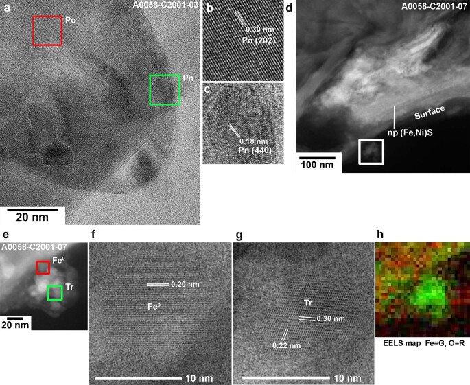
Extended Data Fig. 6. High-resolution images of sulfide and metal in a frothy layer on the large grain A0058–C2001.
(a) A BF-TEM image of a spherical Fe-Ni sulfide bead in the frothy layer in the cross-section sample A0058-C2001–03. (b, c) Enlarged BF-TEM images of the red and green boxed areas are shown in (b) and (c), respectively. The lattice fringes shown in (b) are 0.30 nm, which suggests of pyrrhotite (Po). The fringes shown in (c) are 0.18 nm, suggestive of (4 4 0) of pentlandite (Pn). (d) HAADF-STEM image of the cross-section sample A0058–C2001–07 shows a chain of nanophase (np) (Fe, Ni)S in the thin (~100 nm) smooth layer. (e-h) An aggregate composed of nanophases (<100 nm) on the frothy layer of the cross-section sample A0058–C2001–7. HAADF-STEM image of the aggregate is shown in (e). Enlarged BF-TEM images of the red and green boxed areas in (e) are shown in (f) and (g), respectively. The lattice fringes shown in (b) are 0.30 nm, which suggests of pyrrhotite (Po). The fringes shown in (f) is 0.20 nm, suggestive of (1 1 0) of kamacite (Fe°) and those in (g) are 0.22 and 0.30 nm, suggestive of , and (1 1 0) of troilite (Tr). (h) EELS map of the same area shown in (e) shows that the aggregate lacks oxygen and contains iron.

