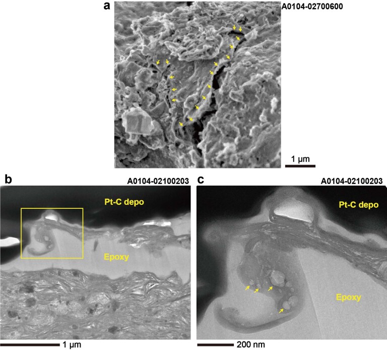
Extended Data Fig. 4. Partially exfoliated smooth layers on phyllosilicate-rich matrix.
(a) Secondary electron image of a smooth layer on the grain A104–02700600. Partial exfoliation of the smooth surface is indicated by yellow arrows. (b-c) BF-TEM image of an exfoliated smooth layer containing vesicles in the cross-section sample A104–02100203. (c)An enlarged image of the area is indicated by a square in (b). The arrows in (c) indicate vesicles in the exfoliated smooth layer. The interstice between the exfoliated layer and the phyllosilicate base is filled by epoxy resin, and Pt-C was deposited by FIB-SEM, both from sample preparation.

