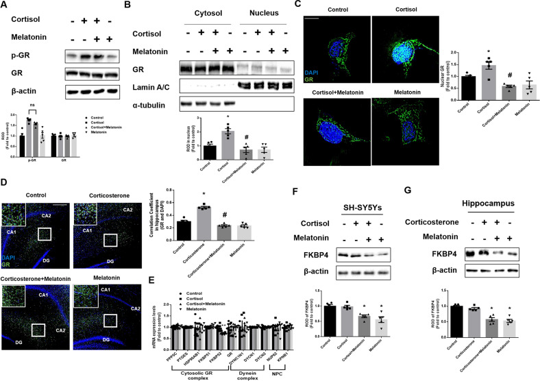
Fig. 3. Melatonin downregulates FKBP4, but is not related to phosphorylation of GR.
A–C, F SH-SY5Y cells were treated with melatonin (1 μM) for 30 min and then with cortisol (1 μM) for 24 h. A The expression of p-GR and GR were detected by western blot. Loading control is β-actin. n = 5. B The expression of GR protein in subcellular fraction samples was detected by western blotting. Lamin A/C and α-tubulin were used as a nuclear and cytosolic loading control, respectively. n = 5. C The cells were immunostained with GR (green) and DAPI (blue). Scale bars, 10 μm (magnification, ×1000). n = 5. D, G Mice were injected with melatonin (10 mg/kg) and then with corticosterone (10 mg/kg) for 7 days. D Slide samples for immunohistochemistry were immunostained with GR (green) and DAPI (blue). Scale bars, 140 μm (magnification, ×100). n = 5. E SH-SY5Y cells were treated with melatonin for 30 min and then with cortisol for 12 h. The mRNA expression of regulatory proteins related to cytosolic GR complex, dynein complex, and NPC were analyzed by real time PCR. n = 5. F, G FKBP4 was detected by western blot. Loading control is β-actin. n = 5. All blots and immunofluorescence images are representative. The representative images were acquired by SRRF imaging system. All data are presented as a mean ± S.E.M. *p < 0.05 versus control, #p < 0.05 versus cortisol or corticosterone.

