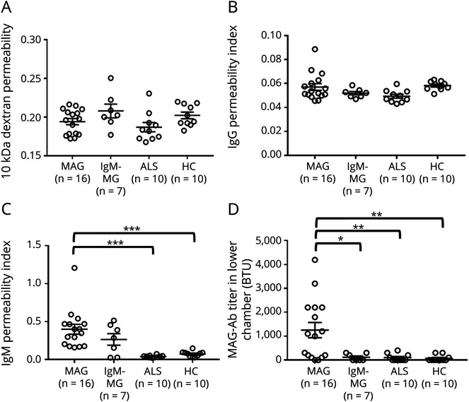
Figure 3. Changes of the 10-kDa Dextran, IgG, IgM, and Anti-MAG Antibody Permeability Evaluated Using an In Vitro Human BNB Coculture Model Exposed to Sera From Individuals With MAG Neuropathy.
PnMECs on the luminal side and human peripheral nerve pericytes on the abluminal side were cultured on a 24-well collagen-coated Transwell culture insert (pore size: 0.4 mm). MCDB 131 medium containing individual 10% sera from patients with anti-MAG neuropathy, patients with IgM-MG neuropathy, patients with ALS, and HCs were incubated with luminal side. The change in the 10-kDa dextran permeability coefficient (A), IgG permeability index (B), IgM permeability index (C), and MAG antibody (MAG-Ab) permeability (D) was calculated. Data are shown as the mean ± SEM. p Values were determined by a one-way ANOVA followed by the Tukey multiple comparison test (*p < 0.05, **p < 0.01, and ***p < 0.001 vs ALS or HC group followed by the Tukey multiple comparison test). ALS = amyotrophic lateral sclerosis; ANOVA = analysis of variance; BNB = blood-nerve barrier; IgM-MG = IgM–monoclonal gammopathy neuropathy; MAG = myelin-associated glycoprotein; PnMECs = peripheral nerve microvascular endothelial cells.

