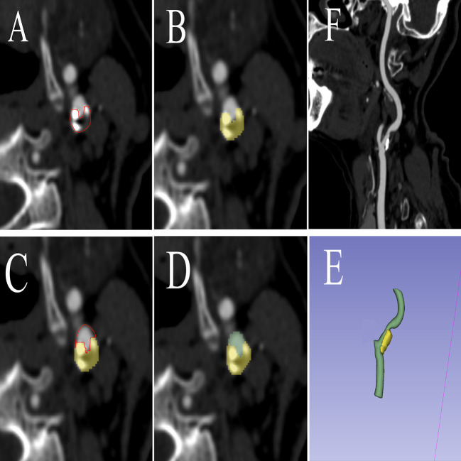
Figure 2.
The procedure of radiomics segmentation. The VOI in each image was segmented with the green arterial lumen and yellow plaques, and a 3D model was synthesized. (A) The edges of atherosclerotic plaques were delineated. (B) Atherosclerotic plaques were segmented. (C) The edges of arteries were delineated. (D) Arteries were segmented. (E) A 3D model was built based on the segmentation of each layer image. (F) Carotid artery CTA corresponding image of the patient. VOI, volume of interest; CTA, computed tomography angiography.

