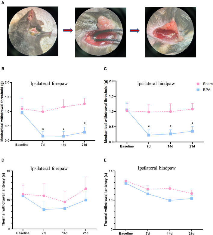
Figure 1.
BPA modeling and pain behavior detection over time. (A) Schematic diagram of BPA operation: from brachial plexus dissociate the C7 nerve separation to nerve avulsion. (B) PMWT results of the operative limb 1 day before and 7, 14, and 21 days after modeling. (C) PMWT results of the ipsilateral hindlimb of sham and BPA mice. (D) No significant difference of TWL in the operative limb between sham and BPA mice at 1 day before and 7, 14, and 21 days after modeling. (E) TWL results of the ipsilateral hindlimb of the two group at each time point. N = 10 per group. *P < 0.05 vs. sham at the same time point. Unpaired T-test was used for data analysis. BPA, brachial plexus avulsion; PMWT, paw mechanical withdrawal threshold; TWL, thermal withdrawal latency.

