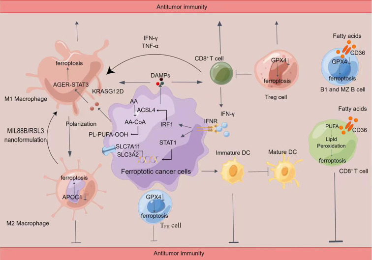
Figure 2.
Crosstalk between immune cells and ferroptosis(By Figdraw)Induction of ferroptosis promotes the release of damage-associated molecular patterns (DAMPs), which in turn activates immune cell activity, including CD8+ T cells and macrophages. Ferroptosis cells significantly cause M1 polarization in macrophages. Ferroptosis can lead to the release of KRASG12D protein from PDAC cells to promote M2 polarization in macrophages via STAT3-dependent fatty acid oxidation. Inhibition of APOC1 or administration of MIL88B/RSL3 nanoformulation promoted the conversion of M2 macrophages into M1 macrophages through ferroptosis. In addition, the release of IFN-γ by CD8+ T cells inhibited the xc (-) system (SLC7A11/SLC3A2) and synergistic-induced ferroptosis in tumor cells through ACSL4, leading to increased sensitivity to ferroptosis. Inhibition of GPX4 caused ferroptosis in Treg cells, TFH cells, B1, and MZB cells. Fatty acids in the tumor microenvironment induce ferroptosis in CD8+ T cells in a CD36-dependent manner. Then impairing the antitumor function of CD8+ T cells. Tumor cells in the early stages of ferroptosis are able to affect the maturation of DC cells and inhibit the phagocytosis of tumor cells by DC cells.

