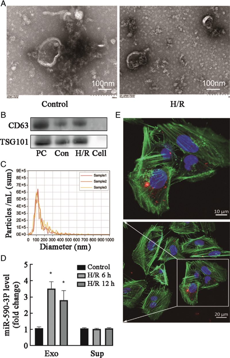
Figure 2.
miR-590-3p is transferred between HK-2 cells via exosomes after H/R treatment. (A) Secretory vesicles isolated from HK-2 cell culture medium were identified using transmission electron microscopy. (B, C) Exosomes were quantified by Western blot and NTA. Cell: Cell supernatant; Con: Control. (D) Levels of miR-590-3p in the exosome (Exo) and supernatant (Sup) fractions of medium from HK-2 cells after 12 h of H/R as detected by real-time reverse transcription-polymerase chain reaction (RT–PCR). Data are expressed as the mean ± standard deviation; N = 3 for NTA, N = 4–5 for Western blot. ∗P < 0.01, vs. the control group. (E) Representative confocal microscopy images of HK-2 cells exposed to PKH26-labeled exosomes from HK-2 cells subjected to 12 h of H/R. Nuclei were stained with DAPI. Red: PKH26; Green: Actin tracker; Blue: DAPI (nuclei). DAPI: 4′,6-diamidino-2-phenylindole; H/R: Hypoxia/reoxygenation; I/R: Ischemia/reperfusion; NTA: Nanoparticle tracking analysis; NC: Negative control; PC: Positive control.

