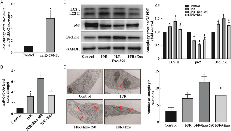
Figure 3.
Exosomal miR-590-3p derived from HK-2 cells after H/R enhanced the activation of autophagy. (A) miR-590-3p expression was significantly upregulated in HK-2 cell exosomes after transfection with a miR-590-3p mimic as determined by RT–PCR. (B) miR-590-3p expression was upregulated in HK-2 cells after H/R and further increased significantly after treatment with miR-590-3p-overexpressing exosomes as determined by RT–PCR. (C) Western blot analysis of autophagy-related proteins (LC3, p62, and Beclin-1) revealed that autophagy was induced in cultured normoxic HK-2 cells after H/R. The activation of autophagy was increased by exosomes overexpressing miR-590-3p from HK-2 cells. (D) Representative images and quantification of autophagic vacuoles in these groups. Autophagic vacuoles (red arrows) were detected by transmission electron microscopy at a magnification of ×10,000. Scale bars represent 2 μm. The data are expressed as the mean ± SD; N = 3 for RT–PCR, N = 4–5 for Western blot. ∗P < 0.05 vs. the control group; †P < 0.05, vs. the H/R group; ‡P < 0.05, vs. the H/R + Exo-590 group. Exo: Exosome; H/R: Hypoxia/reoxygenation; I/R: Ischemia/reperfusion; LC3: Microtubule associated protein 1 light chain 3; SD: Standard deviation.

