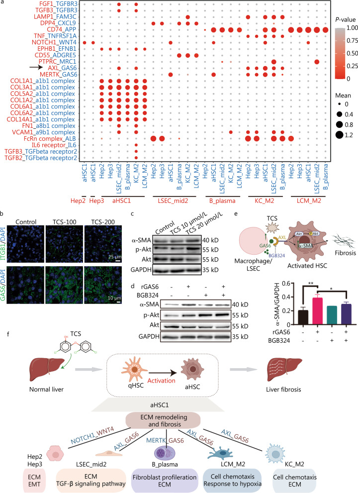
Fig. 7.
Cell–cell communication crosstalk of different cell types. a Dot plot depicting ligand-receptor pairs within different subtypes. Circle sizes indicate mean expression of pairs and colors indicated enrichment of P-values in the two subtypes. b Immunofluorescence staining of ITGB1 (top) and GAS6 (bottom) on mouse liver frozen sections. Scale bar = 25 μm (top) and scale bar = 10 μm (bottom). c LX-2 cells were treated with indicated concentration of TCS for 24 h, and the expression of α-SMA, p-Akt and total Akt were examined with Western blotting, GAPDH was used as loading control. d LX-2 cells were incubated with 500 ng/ml rGAS6 for 30 min and/or pre-incubated with BGB324 (1 μmol/L, 30 min), and the expression of α-SMA, p-Akt and total Akt were examined with Western blotting, GAPDH was used as loading control. e Scheme showing the potential fibrosis-related mechanism caused by the combination of GAS6 and AXL in HSCs. f Summary and inference of cellular communication induced by TCS on mice liver. ITGB1 integrin beta 1, TCS triclosan, α-SMA alpha smooth muscle actin, p-Akt phosphorylated Akt, rGAS6 recombinant GAS6, HSCs hepatic stellate cells, LSEC liver sinusoidal endothelial cells, qHSC quiescent hepatic stellate cells, aHSC activated hepatic stellate cells, ECM extracellular matrix, EMT epithelial-mesenchymal transition; *P < 0.05, **P < 0.01

