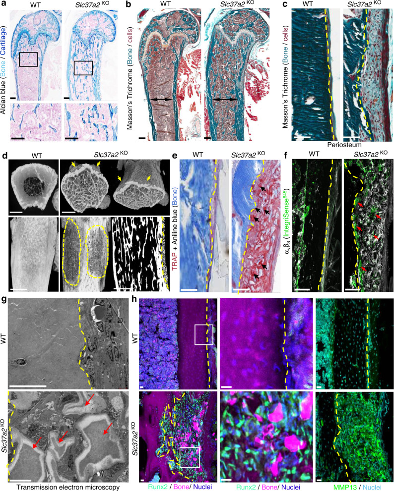
Fig. 6. Slc37a2KO mice exhibit high bone mass and a periosteal bone lesion.
a Representative histological images of Alcian blue staining of the distal femur of 12-week-old female WT and Slc37a2KO mice (n = 5), Bars, 0.1 mm. b and c Mason’s Trichome staining of femurs from 12-week-old female WT and Slc37a2KO mice (b, n = 3) and representative images of the periosteal surface along the metaphyseal–diaphyseal junction (c, n = 3). Black double arrows indicate maximal femoral diameter and dashed lines indicate the periosteal bone surface. Bars, 1 mm. d High-resolution µCT images of 12-week-old female WT and Slc37a2KO femurs indicating the periosteal lesions (arrows and dashed outlines) (n = 2). Bars, 1 mm. e and f Histological evaluation of TRAP+ (e, n = 3) and αvβ3+ (f, n = 3) cells along the periosteal surface of 12-week-old femurs from female WT and Slc37a2KO mice. Dashed lines denote the periosteal bone surface, arrows indicate TRAP+ or αvβ3+ cells. Bars, 1 mm. g Transmission electron microscopic evaluation of the periosteal layer of femurs from WT and Slc37a2KO(n = 2). Dashed yellow lines indicate the periosteal bone surface. Red arrows depict demineralized bone/cartilaginous fragments. Bars, 10 µm. h Representative images of cryosections depicting the periosteal surface along the femurs of 12-week-old female WT and Slc37a2KO mice immunostained for Runx2 or MMP13 (n = 4). Bars, 20 µm.

