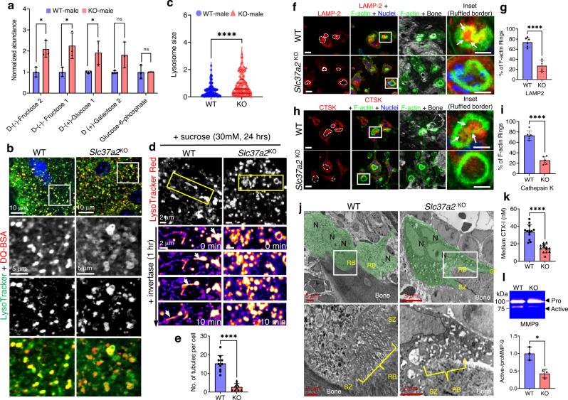
Fig. 9. Export of monosaccharides and delivery of SLs to the ruffled border is impaired Slc37a2KO osteoclasts.
a Quantitative metabolomics profiling of monosaccharide sugars in BMM-derived osteoclasts from WT and Slc37a2KO mice (n = 3; d-(−)-Fructose 2 P = 0.014536; d-(−)-Fructose 1 P = 0.028321; d-(+)-Glucose 1 P = 0.046169). b and c Representative confocal images of WT and Slc37a2KO osteoclasts stained with LysoTracker Green and DQ-BSA (b) and quantitation of SL size (c) (n = 109 SLs pooled from 10 cells per group, P < 0.0001). d Time-lapse confocal image series of WT and Slc37a2KO osteoclasts cultured in high sucrose conditions (30 mM, 24 h) to induce sucrosome formation and imaged for 10 min post-treatment with invertase (0.5 mg/ml, 1 h). SLs were pulsed with LysoTracker Red prior to imaging. Arrows track an SL resolution and tubulation event. e Number of tubules per cell post-treatment with invertase (n = 10 cells, P < 0.0001). f–i Confocal images of WT and Slc37a2KO osteoclasts cultured on bone and immunostained for F-actin in combination with either LAMP-2 (f, Bars, 10 μm) or cathepsin k (CTSK) (h, Bars, 10 μm) and quantitation (g, P < 0.0001; i, P < 0.0001). Arrows indicate delivery of LAMP2 and CTSK within F-actin rings (n = 6). j Representative TEM micrographs of WT and Slc37a2KO osteoclasts (green) lining trabecular bone surfaces within the primary spongiosa of 5-day-old male littermates (n = 3). Magnified pictures illustrate ruffled borders (RB). N nuclei, SZ sealing zone. k, l CTX-1 and active MMP9 levels in media from cultures of bone-resorbing WT and Slc37a2KO osteoclasts as monitored by ELISA (k) and gel zymography (l). n = 13, P < 0.0001 (k) and n = 3, P = 0.0117 (l). Data are presented as means ± SD *P < 0.05 and ****P < 0.0001 by two-tailed unpaired Student’s t-test. Source data are available in the Source Data file. See also related Supplementary Movie 6.

