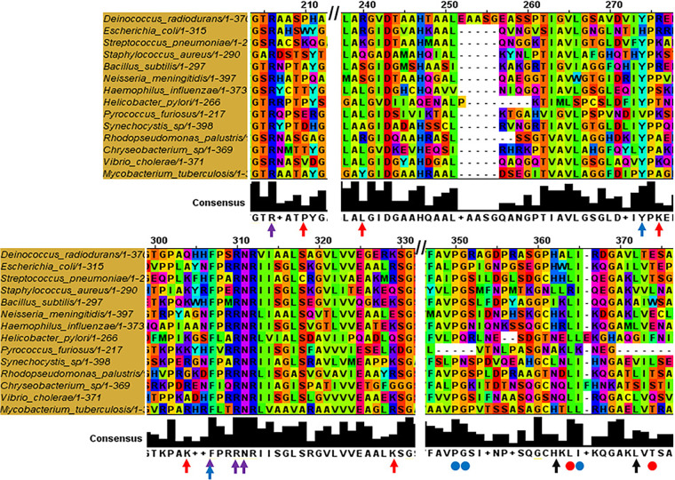
FIG 5.
Sequence alignment of DprAs of different species highlighting conserved residues. Multiple sequence alignment was done using MUSCLE (Multiple Sequence Alignment-EMBL-EBI) and view using Jalview. Red arrow represents conserved lysine (K) of S. pneumoniae (K119, K144, K175, K202, and K225); purple and blue arrows represent conserved H. pylori DprA residues (R52, F-140, R-143, K137, and N-144) responsible for DNA binding; black arrows represent hydrophobic residues of H. pylori DprA (H264, and L273) crucial for dimerization. Red dot represents hydrophobic core dimerizing residues of H. pylori DprA (L196 and F205); blue dots represent the residues involved in self-dimerization of S. pneumoniae DprA (P248, G249, and I263).

