. 2023 Jan 21;14(8):2046–2053. doi: 10.1039/d2sc05848k

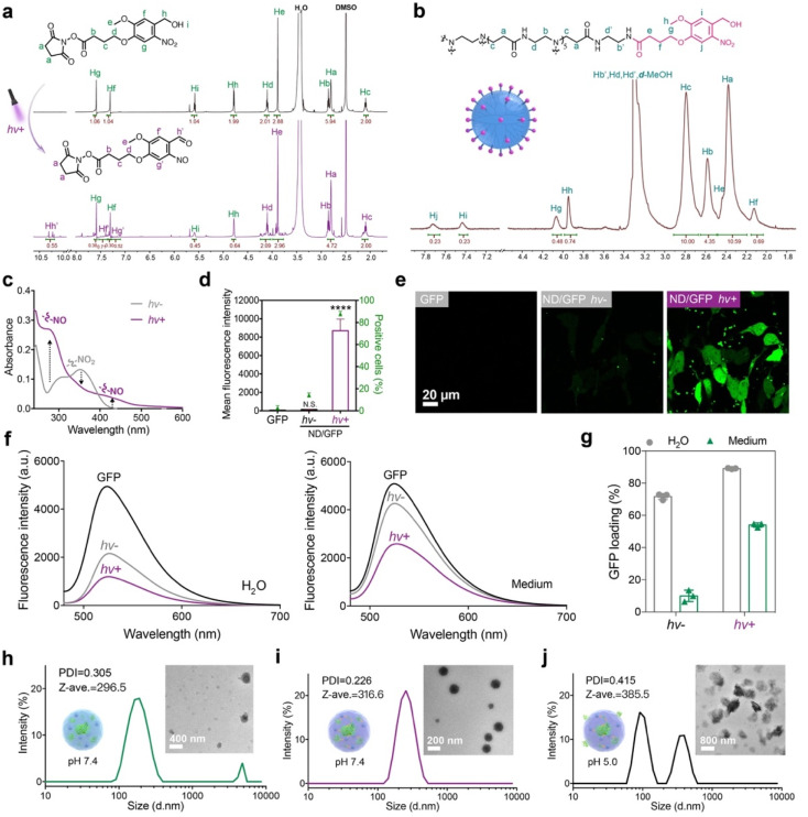
Fig. 2. Photo-activatable properties of the ND polymer in cytosolic protein delivery. (a) 1H NMR spectra of NB-NHS dissolved in DMSO-d6 before and after UV irradiation. (b) 1H NMR spectra of the ND polymer in methanol-d4. (c) UV-vis spectroscopy of the ND polymer before and after UV irradiation for 20 s. (d) Flow cytometry and (e) confocal images of 143B cells treated with ND/GFP complexes with or without UV irradiation. The concentrations of GFP and ND were 16 and 32 μg mL−1, respectively, and the cells were cultured for 24 h before measurement. Data are presented as mean ± standard deviation (s.d.) (n = 3). N.S.p > 0.05 and ****p < 0.0001, calculated by one-way ANOVA. (f) Fluorescence spectra and (g) GFP loading efficiency of the ND/GFP complexes before and after UV light activation in deionized (DI) water and culture media. The concentrations of GFP and ND were 4 and 8 μg mL−1, respectively. DLS and TEM images of ND/GFP complexes without light activation (h), ND/GFP complexes after light activation (i) and the sample of (i) at pH 5.0 (j) in culture media.

Fig. 2