. 2023 Jan 21;14(8):2046–2053. doi: 10.1039/d2sc05848k

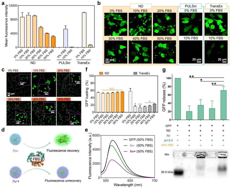
Fig. 3. Serum stability and release responsiveness of the ND polymer in cytosolic protein delivery. Flow cytometry (a) and confocal images (b) of 143B cells treated with the ND/GFP complex after LAC in media containing different contents of FBS for 24 h (GFP, 16 μg mL−1; ND, 32 μg mL−1). Commercial reagents PULSin and TransEx were tested as controls. (c) Confocal images of ND/GFP complexes in culture medium containing different contents of FBS. Schematic illustration about fluorescence change (d) and fluorescence intensity (e) of ND/GFP complexes before and after LAC in culture medium containing 50% FBS. Free GFP was tested as a control. (f) GFP loading efficiency of ND after LAC in culture media containing different contents of FBS (GFP, 4 μg mL−1; ND, 8 μg mL−1). (g) Agarose gel electrophoresis of ND/GFP complexes under different conditions. All data are shown as mean ± s.d. (n = 3). N.S.p > 0.05, *p < 0.01, **p < 0.001, according to one-way ANOVA.

Fig. 3