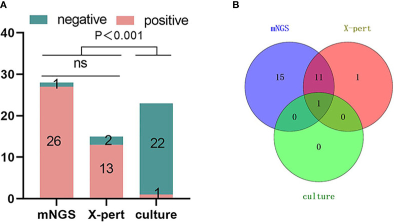
Figure 5.

Comparison of mNGS, X-pert and culture positive rates for the detection of Mycobacterium tuberculosis in spinal tuberculosis focal tissue samples. The number of positive samples detected by the three detection methods showed in (A) that the positive rate of mNGS was higher than that of X-pert, but there was no statistical difference. The positive rate of mNGS and X-Pert was higher than that of culture, and the difference was statistically significant (P < 0.001). (B) Overlapping Venn diagram of samples positive for MTB by the three tests. mNGS, metagenomic next-generation sequencing; X-pert, the X-pert MTB/RIF assay; MTB, Mycobacterium Tuberculosis; ns, no significant difference.
