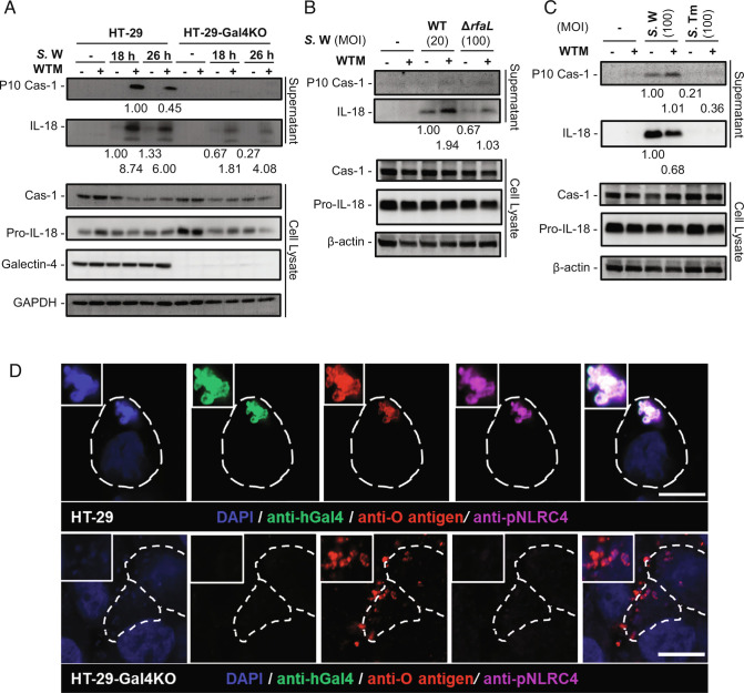
Fig. 4.
Galectin-4 enhances mature IL-18 and caspase-1 (Cas-1) production from epithelial cells after bacterial infection especially following autophagy inhibition. (A) HT-29 and HT-29-Gal4KO cells treated with or without wortmannin (WTM) were infected with S. enterica serovar Worthington (S.W), MOI = 100. Supernatants and cell lysates were collected 18 and 26 h post-infection and analyzed by immunoblotting with the indicated antibodies. (B) HT-29 cells treated with or without WTM were infected with WT (MOI = 20) or ΔrfaL (MOI = 100) S.W. Supernatants and cell lysates were collected 24 h post-infection and analyzed by immunoblotting with the indicated antibodies. (C) HT-29 cells treated with or without WTM were infected with S.W or S. enterica serovar Typhimurium (S.Tm), MOI = 100. Supernatants and cell lysates were collected 24 h post-infection and analyzed by immunoblotting with the indicated antibodies. The membranes for supernatants in (A–C) were stripped and reprobed for IL-18 after initial probing for Cas-1. The relative amounts of P10 Cas-1 to Cas-1 and IL-18 to pro-IL-18, as determined by densitometric analysis, are indicated. The membrane for lysates in (A) was stripped and reprobed for GAPDH, Pro-IL-18, and then galectin-4 after initial probing for Cas-1. The membranes for lysates in (B and C) were stripped and reprobed for Pro-IL-18, and then β-actin after initial probing for Cas-1. In (A–C) the data are representative of the results from two independent experiments. (D) HT-29 and HT-29-Gal4KO cells treated with WTM were infected with S.W, MOI = 100. Confocal images of cells stained with S.W. DAPI (blue), anti-human galectin-4 (anti-hGal4, green), anti-O antigen of Salmonella (anti-O antigen, red), and anti-phospho-NLRC4 (anti-pNLRC4, purple). The borders of the infected HT-29 cells are outlined with white dashed lines. (Scale bars, 10 µm.)

