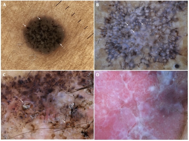
Figure 2.

Examples of skin tumors in dark-skinned patients (phototypes V/VI) typified by the newly-introduced dermoscopic structures: black, small clods (black globules) in a seborrheic keratosis (arrows) (a); follicular plugs in an actinic keratosis (arrows) (b); erosions in a basal cell carcinoma (arrows) (c); and white color around vessels (perivascular white halo) in a squamous cell carcinoma (d).
