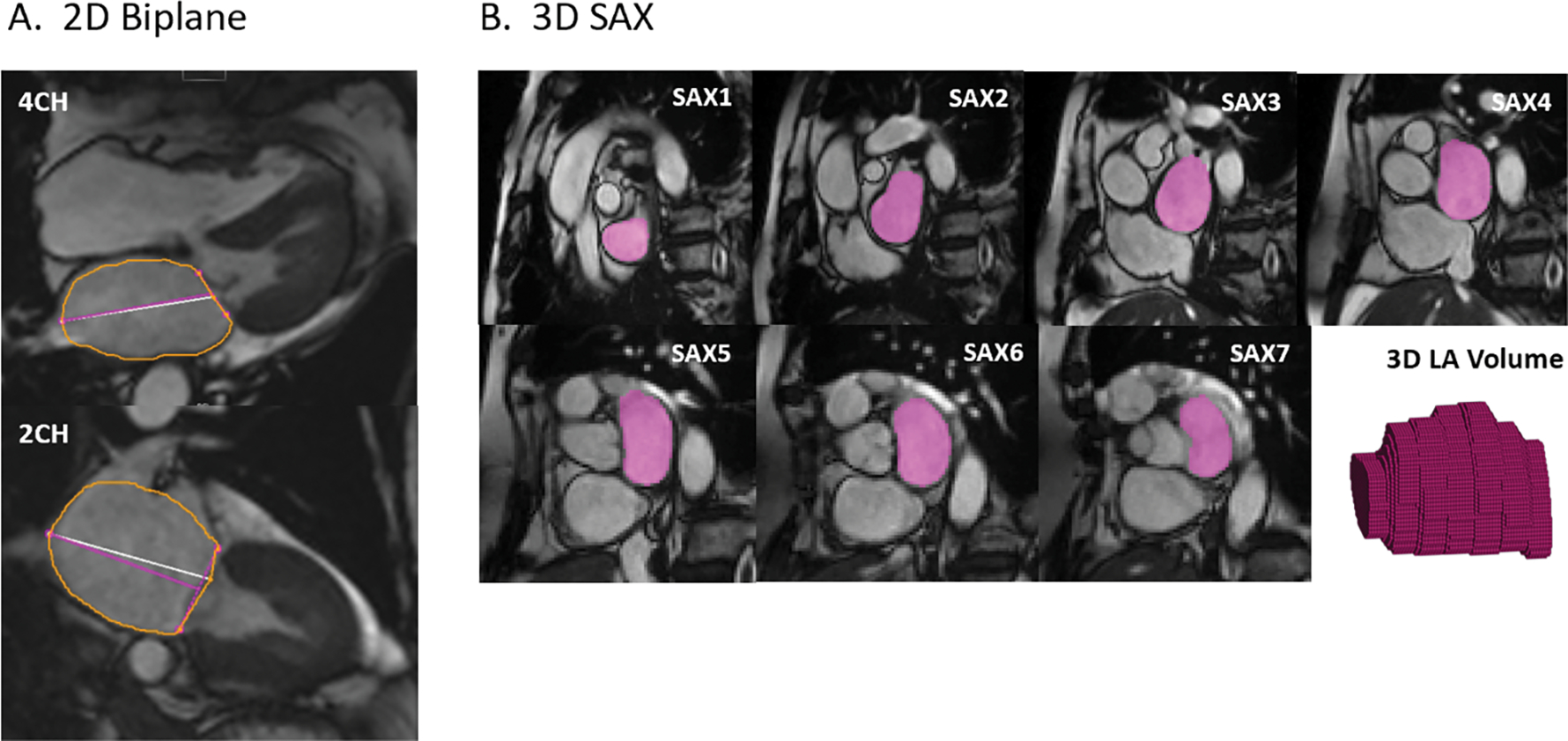
Figure 2.

(A) Biplane method: LA endocardial boundaries were contoured on two- and four-chamber images at max LAV. (B) 3D assessment: Contours on SAX images and a 3D volume representation of the LA endocardium for a single time point; the contouring was performed for each time point of the cardiac cycle (25 per patient). At the atrioventricular border, the LA was contoured if less than 50% of left ventricular myocardium was visible.
