
Figure 1.
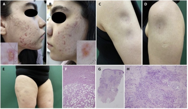
(A,B) Multiple bizarre cicatricial acneiform lesions on cheeks with multiple linear and branching vessels over translucent yellowish-orange globular structure on scar-like areas, dermoscopically. (C) Multiple subcutaneous atrophic nodular lesions on the right arm. (D) Subcutaneous atrophic nodular lesion on the left arm. (E) Multiple subcutaneous nodular lesions on the lower proximal extremity. (F) Septal thickening and lobular inflammation rich in histiocytes (H&E, ×200). (G,H) Periadnexial inflammation with focal granulomatous reaction (H&E, ×40, ×200).